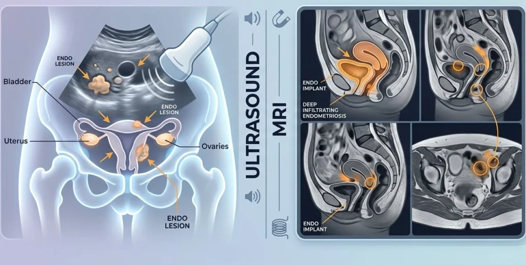
Thousands of women worldwide struggle with endometriosis, a chronic medical condition that can contribute to pelvic pain, inflammation, and fertility problems. Obtaining an accurate diagnosis can sometimes be difficult because its symptoms may overlap with those of other pelvic conditions. Endometriosis radiology plays an important role in this process.
Medical professionals can use advanced imaging techniques to identify certain forms of endometriosis, assess the extent of the disease, and support individualized treatment planning. Ultrasound and MRI have become particularly valuable for identifying ovarian endometriomas and deep endometriosis, although imaging cannot detect every type of endometriosis.
This blog discusses the imaging techniques used in the evaluation of endometriosis, how radiology helps with detection and treatment planning, and why expert guidance is important for effective management.
What is Endometriosis?
Endometriosis is a chronic condition in which tissue similar to the lining of the uterus (endometrium) grows outside the uterus.
It can commonly affect:
- The ovaries
- The fallopian tubes
- The peritoneum and other areas of the pelvis
- The uterosacral ligaments and other pelvic structures
- The bowel
- The bladder and urinary tract in some cases
These endometriosis lesions can respond to hormonal changes during the menstrual cycle and may contribute to inflammation, pain, adhesions, and scarring. The condition can also affect fertility in some women.
Because symptoms can vary considerably from one person to another, imaging has become an important part of the diagnostic work-up.
Endometriosis Radiology
Endometriosis radiology refers to the use of medical imaging techniques to help identify endometriosis, assess its location and extent, and support treatment planning.
Endometriosis imaging can help specialists to:
- Identify ovarian endometriomas
- Detect deep endometriosis
- Assess the involvement of surrounding pelvic organs
- Evaluate the extent of disease before surgery
- Support fertility and treatment planning
Current guidelines recommend imaging, particularly ultrasound or MRI, as part of the diagnostic work-up for suspected endometriosis. However, a normal scan does not completely exclude the condition, particularly superficial endometriosis.
Why Does Radiology Matter?
Many women experience symptoms that may resemble those of other gynecological or pelvic conditions. This can sometimes contribute to delays in diagnosis.
Radiology can help with:
- Identifying structural abnormalities
- Detecting ovarian endometriomas
- Identifying deep pelvic endometriosis
- Assessing the involvement of nearby organs
- Supporting treatment and surgical planning
Imaging findings can provide valuable information to gynecologists and fertility specialists when they are considered alongside symptoms, medical history, and clinical examination.
Endometriosis and Ultrasound
For women suspected of having endometriosis, transvaginal ultrasound is commonly used as an initial imaging investigation. A small ultrasound probe is inserted into the vagina to produce detailed images of the pelvic organs.
Ultrasound is particularly useful for identifying:
- Ovarian endometriomas
- Some forms of deep endometriosis
- Pelvic anatomical abnormalities
- Changes involving the uterus and ovaries
A specialized transvaginal ultrasound performed by an experienced examiner can be particularly useful when deep endometriosis is suspected.
However, small or superficial endometriotic lesions may not be visible on ultrasound. Therefore, a normal ultrasound does not necessarily mean that endometriosis is absent.
MRI for Endometriosis
When deep endometriosis is suspected, Magnetic Resonance Imaging (MRI) can provide detailed images of soft tissues and the pelvic organs.
MRI can help evaluate:
- The uterus
- The ovaries
- The bowel
- The bladder
- The pelvic ligaments
- The rectovaginal region
- Other areas that may be affected by deep endometriosis
MRI can be particularly helpful for assessing the location and extent of deep endometriosis and for planning complex surgery. Depending on the patient’s symptoms and the findings on ultrasound, a specialist may recommend MRI as part of the preoperative evaluation.
Common Radiological Findings
Radiologists may identify several findings associated with endometriosis.
1. Ovarian Endometriomas
Ovarian endometriomas, sometimes called “chocolate cysts,” are cysts in the ovary associated with endometriosis and containing altered or old blood.
Imaging may show characteristic features such as:
- Homogeneous, low-level internal echoes on ultrasound
- Thickened cyst walls in some cases
- Characteristic signal patterns on MRI, including high signal intensity on T1-weighted images and low signal intensity on T2-weighted images
These imaging features can help specialists distinguish endometriomas from other types of ovarian cysts.
2. Deep Infiltrating Endometriosis
Deep endometriosis occurs when endometriotic lesions extend deeply into tissues and may involve structures such as:
- Uterosacral ligaments
- Rectovaginal region
- Bowel
- Bladder
- Other pelvic structures
Specialized ultrasound and MRI can help identify these lesions and assess their extent. This information can be particularly valuable when surgery is being considered because it may help the surgical team plan the appropriate approach.
3. Adhesions
Adhesions are bands of scar-like tissue that can develop as a result of inflammation and previous disease or surgery. Endometriosis can be associated with pelvic adhesions.
Adhesions themselves may not always be directly visible on imaging. However, radiologists may identify indirect signs, such as restricted movement between pelvic organs, abnormal anatomy, or organs appearing fixed in an unusual position.
Can Every Case of Endometriosis Be Diagnosed by Radiology?
No.
One of the major limitations of imaging is that very small or superficial endometriotic lesions may not be visible on ultrasound or MRI. A woman can therefore have endometriosis even when imaging results appear normal.
For this reason, specialists consider several factors when evaluating suspected endometriosis, including:
- Signs and symptoms
- Medical history
- Physical examination
- Imaging findings
- Response to treatment
- Fertility concerns
If symptoms continue despite negative imaging, or if empirical treatment is unsuccessful or inappropriate, laparoscopy may be considered in selected patients. Current guidelines emphasize that a normal ultrasound or MRI does not automatically rule out endometriosis.
Assessment of Fertility and Radiology
Imaging can provide important information when endometriosis is being evaluated in women who are experiencing fertility problems.
Radiology can help assess:
- Ovarian endometriomas
- The structure and anatomy of the pelvis
- Uterine abnormalities
- The presence and extent of endometriosis
- Findings that may be relevant to fertility treatment or surgery
Ultrasound can also be used during fertility treatment to monitor ovarian follicles and ovulation. However, ovarian reserve is assessed using a combination of clinical evaluation, ovarian reserve tests, and ultrasound-based measurements such as antral follicle count rather than imaging alone.
The information obtained through these assessments can help fertility specialists develop individualized treatment plans.
When is Imaging Suggested?
A specialist may recommend imaging when a woman experiences symptoms or has clinical concerns such as:
- Severe or persistent pelvic pain, particularly around menstruation
- Pain during sexual intercourse
- Heavy or painful menstrual bleeding
- Difficulty getting pregnant
- Suspected ovarian endometrioma
- Symptoms suggesting deep endometriosis
The choice of imaging depends on the patient’s symptoms, examination findings, and the type of endometriosis being suspected.
Preparing Yourself for an Endometriosis Scan
Preparation depends on the type of imaging examination being performed.
Patients may be advised to:
- Arrive with a full bladder for certain ultrasound examinations
- Follow specific preparation instructions before an MRI, if required
- Bring previous imaging reports and relevant medical records
- Inform the radiologist or imaging team about symptoms, previous surgeries, and relevant medical history
- Follow any dietary or medication instructions provided by the imaging center
Following the recommended preparation instructions can help ensure that the examination is performed effectively.
Advanced Endometriosis Imaging at Top Fertility Care
Proper treatment begins with a precise diagnosis. Women suspected to have endometriosis may undergo complete reproductive evaluations supported by advanced imaging and fertility assessments at NJINSKY IVF & Fertility Clinic.
This clinic assesses pelvic pain, cysts in the ovaries, and fertility issues by integrating the knowledge of gynecologists, fertility experts, and experienced radiologists.
NJINSKY IVF & Fertility Clinic offers fertility treatment services like ovulation monitoring, hormonal evaluations, IVF, IUI, and embryo transfer in addition to diagnostic imaging.
This clinic forms treatment strategies that target both the management of symptoms and potential fertility goals by merging advanced reproductive healthcare with modern radiological imaging.
Concluding Remarks
The use of modern imaging has changed the way medical professionals evaluate women with persistent pelvic pain and fertility concerns related to endometriosis. Ultrasound and MRI can provide important information about ovarian endometriomas, deep endometriosis, pelvic anatomy, and the extent of disease, even though imaging cannot detect every case.
Radiology specialists and gynecologists can make better-informed decisions about treatment, surgery, and fertility planning when imaging findings are considered alongside a thorough clinical evaluation. Women who experience ongoing symptoms should consult a qualified gynecologist or appropriate specialist, since timely evaluation and individualized treatment can help improve symptom management, quality of life, and reproductive care.